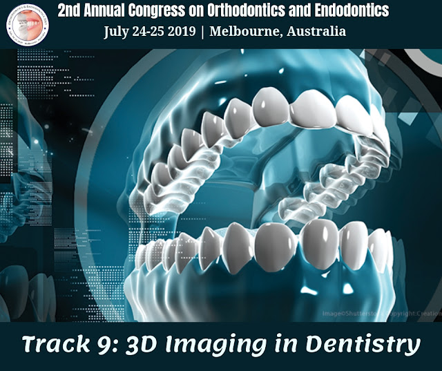
Track 9: 3D Imaging in Dentistry
3D dental imaging is one of the most important and exciting developments in dental diagnostic technology in the past decade. Embed medical procedure, similarly as with any restorative or dental intercession, requires watchful wanting to guarantee no coincidental harm is caused. 3D imaging enables us to take a gander at your jawbone from all edges on a PC screen and survey painstakingly what careful alternatives are accessible to you. 3D imaging has been broadly utilized in different fields of dentistry to help determine, in treatment arranging and machine development. CBCT Technology is the most up to date advancement in 3D imaging. It utilizes comparative innovation as customary CT scanners, however, can do it with less radiation than a conventional arrangement of dental x-beams. It can give a top-notch three-dimensional picture inside seconds.
- Produce more accurate images
- Assist in dental implant placement
- Help our dentists recognize serious problems earlier
- Allow us to share information with other healthcare professionals, dentists, and insurance agencies, as needed
Advantages:
A dental 3D scan allows clinicians to view dental anatomy from different angles. A 3D scan can help gain a better view of bone structures, such as adjacent root positions, in order to locate canals and root fractures, as well as provide the ability to more accurately measure anatomical structures. These scans also support a wide range of diagnosis and treatment planning, making them extremely flexible. Further, they increase the possibility of treatment success, granting practitioners greater predictability and confidence in preparing for extractions, performing root evaluations, and placing implants.
3D dental imaging also delivers the power of repeatability, providing fast and accurate imaging that’s consistent—and thus, reliable. Using a 3D dental scanner equips dental professionals with a comprehensive view, letting them see specific conditions in the region of interest to determine whether a treatment is necessary. Because details show up so clearly, patients can be more confident in a dentist’s decision. In addition, the use of dental imaging technology often creates a more comfortable and engaging dental visit for the patient.
The Gendex GXDP-700 Series features the pinnacle of 3D dental imaging technology, allowing dentists to plan for more predictable treatment outcomes by taking advantage of powerful 3D software analysis and simulation tools. Plus, dental practitioners can control the exposure and the size of scanned areas using the system’s flexible field-of-view (FOV) to meet individual patient and clinical needs. As a practice grows to offer additional imaging capabilities, the GXDP-700 imaging solution can be upgraded within your own timeline and budget.
X-ray imaging, including dental 3D (CBCT), provides a fast, non-invasive way of answering a number of clinical questions. Dental CBCT images provide three-dimensional (3D) information, rather than the two-dimensional (2D) information provided by a conventional X-ray image. This may help with the diagnosis, treatment planning and evaluation of certain conditions. Dental CBCT should be performed only when necessary to provide clinical information that cannot be provided using other imaging modalities. Concerns about radiation exposure are greater for younger patients because they are more sensitive to radiation. For more information about the use, benefits, and risks of CBCT
Disadvantages:
Costs– the initial costs associated with digital equipment is one major disadvantage with dental practices. To purchase the computer hardware and software, and the digital imaging sensors can be costly. And just like dental equipment, there is maintenance, repairs, software updates, and equipment replacement that must be considered when changing from radiographic film to digital imaging. Just as it is with radiographic film, digital imaging will require providers to think about asepsis or infection control when they’re taking images. The practice will need to purchase disposable barriers to be placed over phosphor plates and hard sensors, as the sensors cannot be sterilized. Phosphor plates can be reused a number of times before they need to be replaced. However, the damage will require phosphor plates be replaced more often. Damaged phosphor plates can obscure the image not allowing the dentist to make thorough diagnoses.
Patient Comfort– direct sensors are hard and not flexible like film. They can be rather thick, depending on the manufacturer. However, some of the hard sensors on the market have excellent contrast and density, allowing for enhanced diagnoses of incipient lesions. In fact, just in the last few years, the diagnostic value has increased considerably due to increased technology of hard sensors. However, patient comfort and limited positioning of the sensor intraorally due to patient anatomy is one disadvantage that prohibits some dental practices from considering purchasing hard sensors. With all types of digital sensors, training will be needed to show dental providers how to place sensors to allow for the most coverage of anatomy.
Medico-legal– concerns regarding manipulating images has been addressed by manufacturers. Dental software has warning features if the enhanced image does not match the original image. It is recommended copies of the original images be stored on the computer or network server. Researchers in the area of fraudulent digital dental records recommend digital content have attached metadata. In forensic dentistry metadata and watermarking be added to discourage tampering documents.
Share your research on Intraoral Digital Imaging at our 2nd Annual Congress on Orthodontics and Endodontics to be held in Melbourne, Australia from July 24-25 2019
Submit Your Research (Click Here)
Register Now (Click Here)


In Raleigh, the provision of dental services is beginning to change, thanks to the progressive thinking of some dentist raleigh. This change involves making a broader range of dental care services available under a single roof.
ReplyDelete